Lesson 12: Healing Progression: Laser Burn on Tanned Skin
Six-week documentation of a laser burn that occurred when treatment was performed over recently
tanned skin. Excess surface pigment absorbed the energy, causing thermal injury. This series shows
the full healing arc from acute injury to resolution.
Week 1
Acute injury shortly after treatment. Oval burn pattern corresponding to the laser spot size is visible across the treatment area. Surrounding redness and inflammation are prominent.
Image used with client consent for educational purposes. © Knue Laser.
Week 2
Early breakdown of the burn sites. Blistering, surface disruption, and oozing are visible at the deepest points of thermal injury.
Image used with client consent for educational purposes. © Knue Laser.
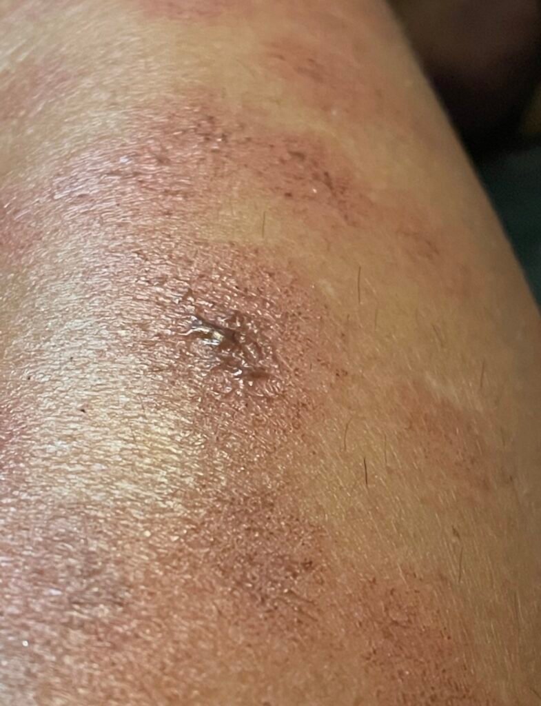
Week 3
Wounds remain open and inflamed. Wound care and consulting physician involvement are required at this stage. Risk of secondary infection is elevated.
Image used with client consent for educational purposes. © Knue Laser
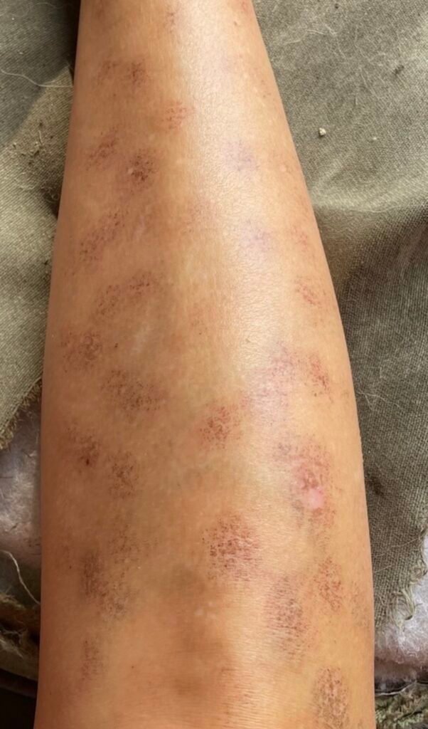
Week 4
Transition out of the acute phase. Scabbing has formed at injury sites and surface tissue is beginning to repair beneath the crust.
Image used with client consent for educational purposes. © Knue Laser
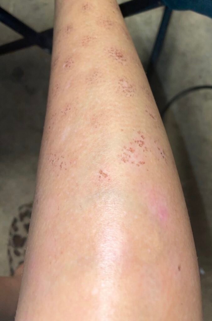
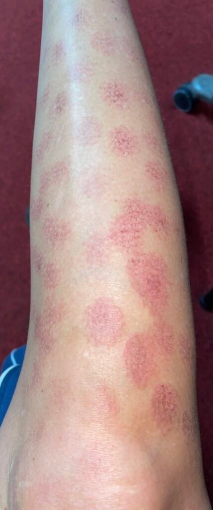
Week 5
Surface lesions are resolving. Post-inflammatory hyperpigmentation is developing at the injury sites as the acute inflammation subsides.
Image used with client consent for educational purposes. © Knue Laser.
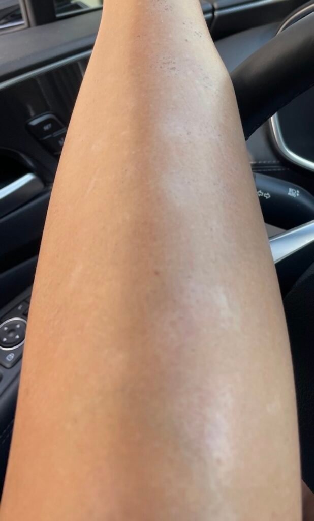
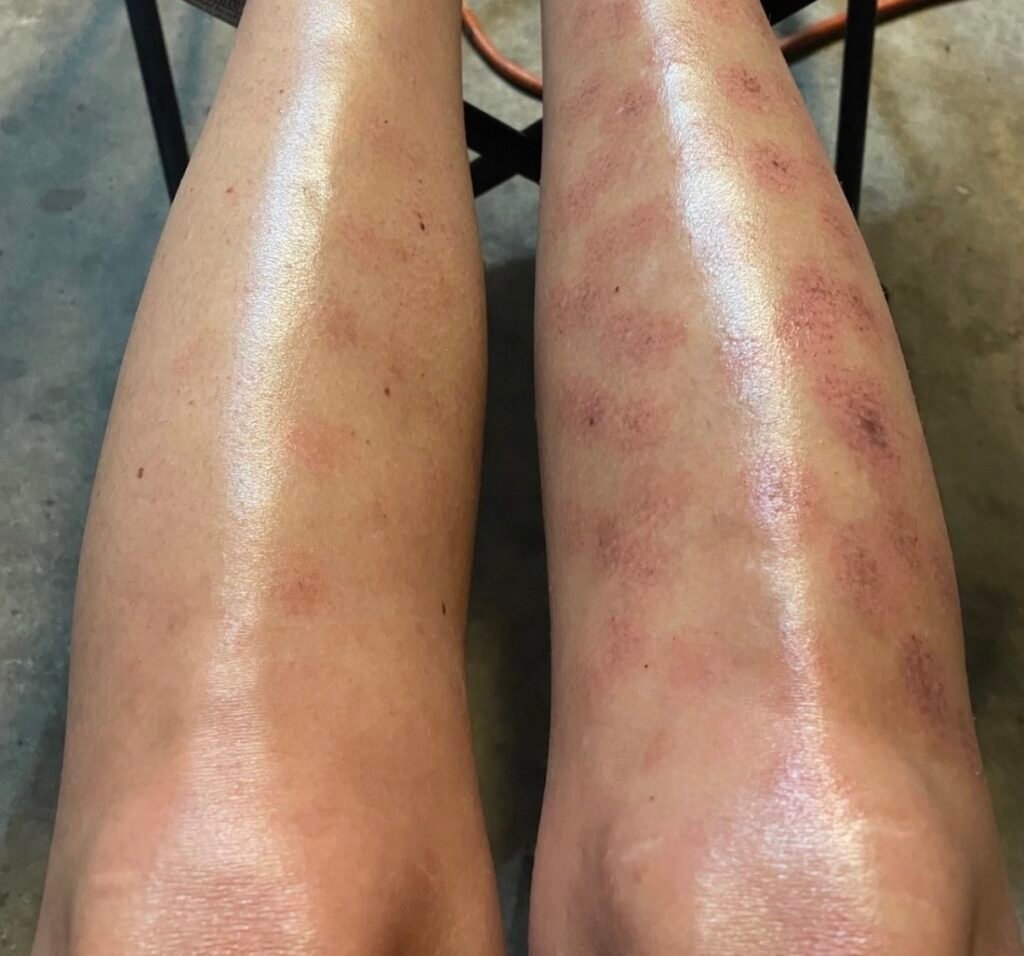
Week 6
Late healing phase. Surface has closed but residual pigment changes and textural differences remain. Scarring and long-term pigment changes are possible outcomes, particularly when care is delayed or the injury is deep.
Image used with client consent for educational purposes. © Knue Laser.